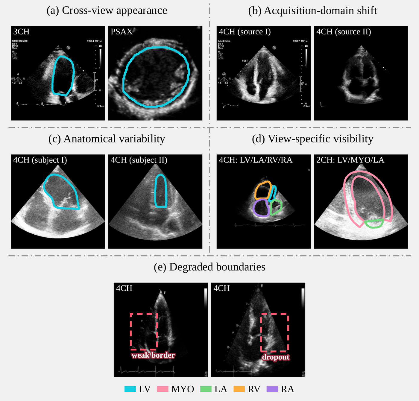
Why one model is hard
✨ arXiv preprint, 2026 ✨
*Equal contribution
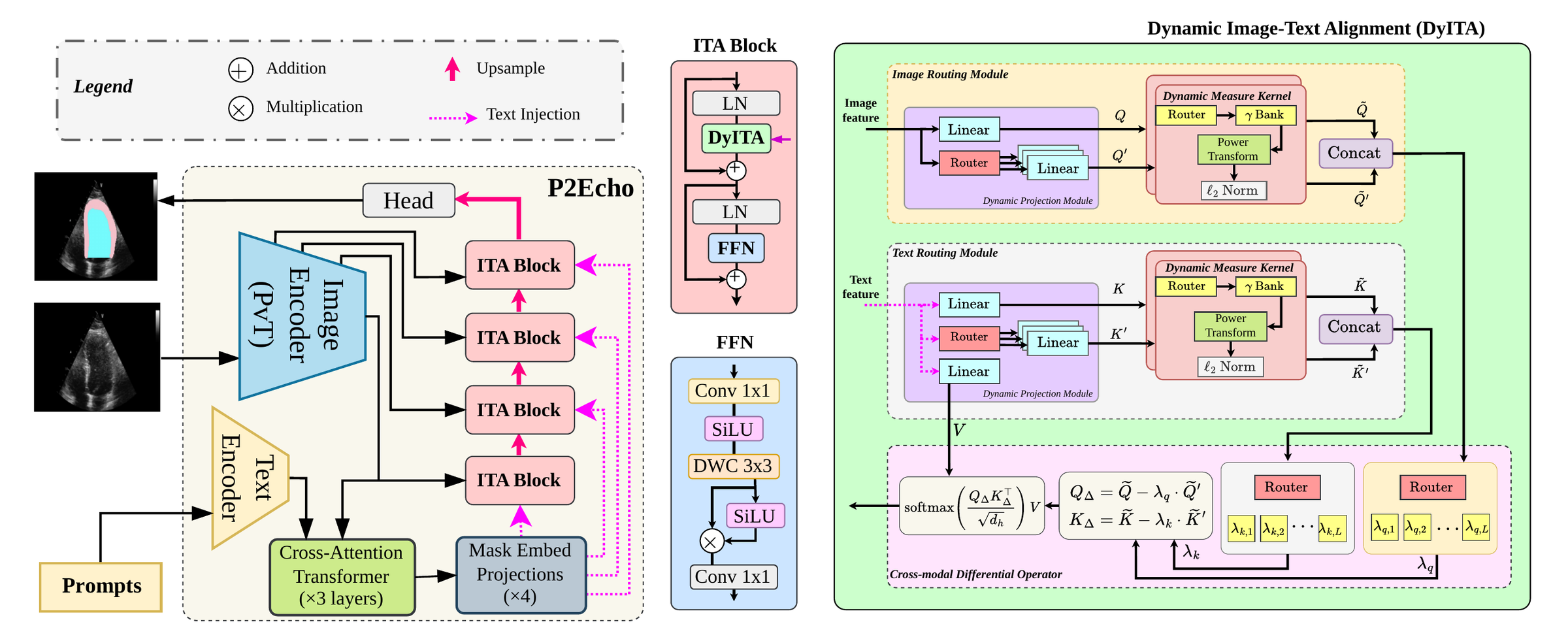
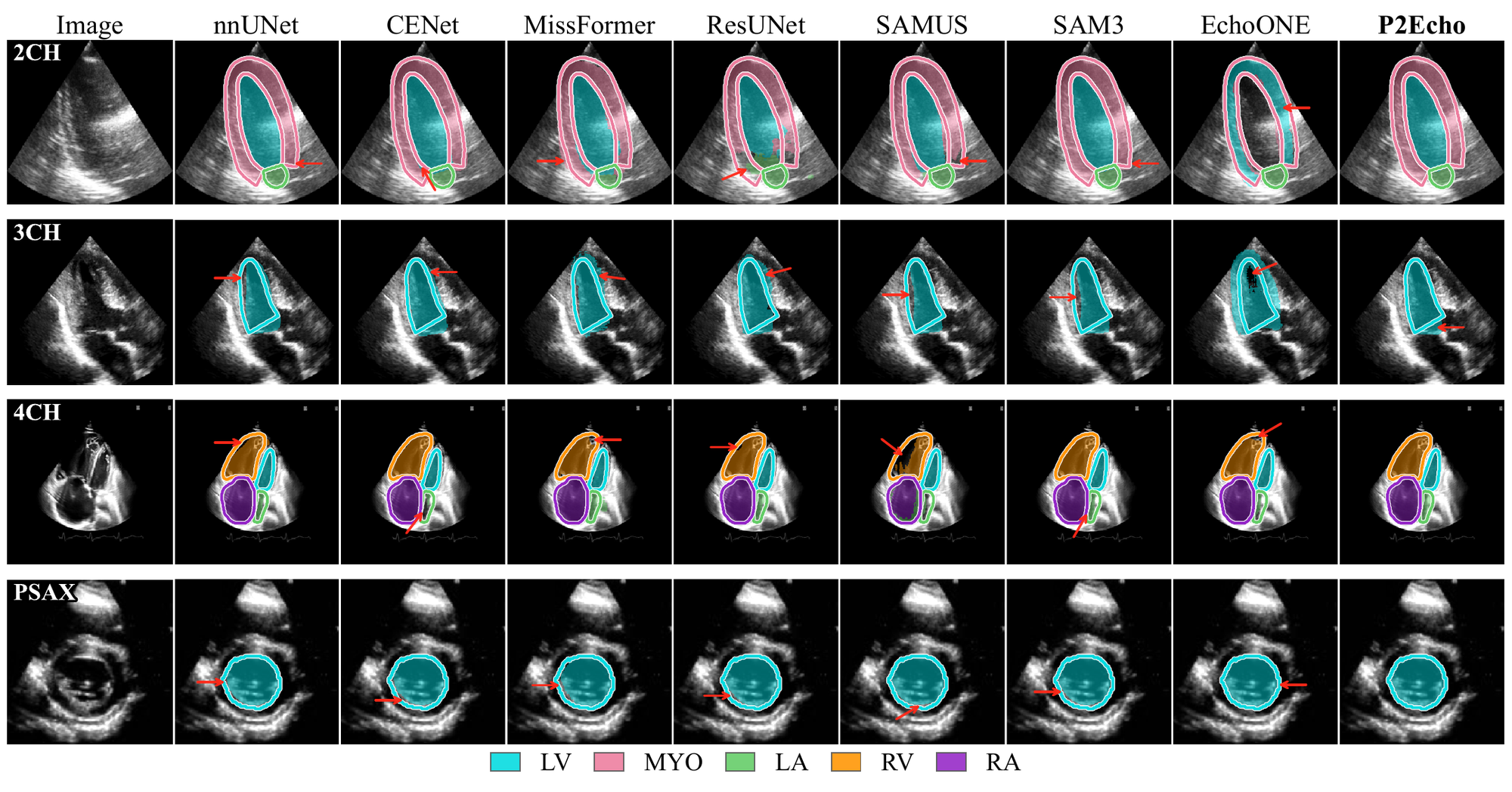
Automated segmentation in multi-view echocardiography is essential for comprehensive cardiac assessment, yet remains challenging due to substantial anatomical and appearance variability across standard acoustic windows. In this paper, we present P2Echo, a unified framework capable of segmenting diverse cardiac structures across multiple standard echocardiographic views using a single model controlled by structured view-aware text prompts. Unlike recent foundation model adaptations that rely on implicit latent priors, P2Echo explicitly conditions the segmentation process via a novel Dynamic Image-Text Alignment (DyITA) module. DyITA integrates a differentiable routing mechanism and token differential operators to dynamically modulate decoder features according to the requested view and target structure. This allows the network to adaptively switch its processing pathways for different views without architectural changes. Extensive experiments on a large-scale internal dataset (seen during training) and held-out evaluation on three external datasets demonstrate that P2Echo matches or exceeds both view-specific specialists and state-of-the-art universal models at a fraction of their computational cost, offering an efficient, controllable solution for multi-plane cardiac analysis. Code is available at github.com/moeinheidari7829/P2Echo.
| Method | 2CH | 3CH | 4CH | PSAX | ||||||||
| mDice ↑ | mIoU ↑ | HD95 ↓ | mDice ↑ | mIoU ↑ | HD95 ↓ | mDice ↑ | mIoU ↑ | HD95 ↓ | mDice ↑ | mIoU ↑ | HD95 ↓ | |
| nnUNet | 92.82 ± 3.75 | 86.82 ± 6.14 | 4.90 ± 2.20 | 93.07 ± 2.97 | 87.17 ± 5.03 | 5.98 ± 3.02 | 91.39 ± 5.68 | 84.64 ± 8.17 | 6.35 ± 4.02 | 91.50 ± 5.69 | 84.78 ± 8.54 | 7.72 ± 4.86 |
| SwinUNet | 86.58 ± 6.08 | 77.15 ± 7.93 | 7.75 ± 3.76 | 87.27 ± 6.03 | 77.88 ± 8.89 | 8.86 ± 4.97 | 85.43 ± 8.16 | 75.62 ± 10.04 | 9.04 ± 6.51 | 85.29 ± 8.96 | 75.29 ± 12.06 | 11.11 ± 7.10 |
| ResUNet | 91.80 ± 2.89 | 85.21 ± 4.53 | 5.46 ± 3.30 | 91.78 ± 3.60 | 85.26 ± 5.81 | 5.69 ± 4.01 | 88.09 ± 6.52 | 80.01 ± 8.57 | 7.83 ± 10.28 | 88.99 ± 7.45 | 80.86 ± 10.67 | 14.88 ± 15.12 |
| MissFormer | 90.88 ± 3.04 | 83.70 ± 4.73 | 5.48 ± 1.62 | 91.09 ± 5.65 | 84.12 ± 8.43 | 5.95 ± 4.07 | 88.34 ± 5.67 | 79.92 ± 7.72 | 6.24 ± 4.16 | 88.50 ± 7.24 | 80.00 ± 10.26 | 9.20 ± 5.19 |
| CENet | 93.11 ± 2.14 | 87.38 ± 3.60 | 4.07 ± 1.25 | 93.09 ± 2.99 | 87.42 ± 5.03 | 4.42 ± 2.10 | 90.88 ± 4.80 | 83.89 ± 6.56 | 4.71 ± 3.02 | 91.08 ± 5.10 | 83.99 ± 8.02 | 7.29 ± 3.46 |
| SAMUS | 89.04 ± 10.39 | 81.85 ± 12.13 | 13.67 ± 3.38 | 93.61 ± 2.91 | 88.13 ± 4.97 | 8.42 ± 1.18 | 89.36 ± 9.15 | 82.00 ± 11.44 | 13.72 ± 3.47 | 91.46 ± 4.23 | 84.55 ± 6.86 | 12.28 ± 2.17 |
| SAM3 (Zero-shot) | 8.75 ± 6.41 | 4.84 ± 4.23 | 220.04 ± 7.70 | 2.37 ± 1.34 | 1.29 ± 0.26 | 310.12 ± 20.45 | 6.32 ± 4.22 | 3.53 ± 2.01 | 241.67 ± 30.12 | 5.06 ± 2.66 | 2.99 ± 2.20 | 306.86 ± 11.85 |
| SAM3 (Fine-tuned) | 93.39 ± 1.75 | 87.60 ± 3.11 | 3.18 ± 0.89 | 92.70 ± 2.48 | 87.04 ± 4.21 | 6.69 ± 1.70 | 90.85 ± 5.25 | 83.85 ± 7.59 | 4.19 ± 3.53 | 91.18 ± 4.66 | 84.13 ± 7.32 | 7.90 ± 3.85 |
| MedCLIP-SAMv2 | 28.98 ± 16.00 | 17.98 ± 11.14 | 170.40 ± 79.67 | 31.70 ± 12.76 | 19.48 ± 8.57 | 170.09 ± 60.78 | 24.61 ± 14.78 | 14.87 ± 10.07 | 170.90 ± 86.40 | 27.72 ± 15.34 | 17.04 ± 10.72 | 36.48 ± 10.60 |
| Grounding DINO-US-SAM | 73.97 ± 21.04 | 62.20 ± 21.16 | 15.47 ± 19.39 | 83.29 ± 7.09 | 71.92 ± 9.60 | 10.59 ± 5.61 | 76.43 ± 13.88 | 64.30 ± 16.02 | 12.31 ± 7.41 | 84.42 ± 9.54 | 74.06 ± 12.55 | 13.50 ± 8.19 |
| EchoONE | 91.69 ± 11.67 | 85.83 ± 12.64 | 2.51 ± 3.38 | 93.60 ± 3.35 | 88.15 ± 5.48 | 4.24 ± 2.19 | 91.11 ± 10.10 | 84.69 ± 11.62 | 3.88 ± 3.50 | 91.71 ± 8.82 | 85.56 ± 10.70 | 7.97 ± 3.06 |
| P2Echo (ours) | 96.94** ± 1.72 | 94.11** ± 3.08 | 2.03** ± 0.92 | 94.71** ± 2.16 | 90.03** ± 3.79 | 3.96** ± 1.60 | 93.87** ± 5.07 | 88.81** ± 7.48 | 3.33** ± 3.77 | 92.00 ± 4.97 | 85.52 ± 7.76 | 6.99 ± 3.74 |
| Method | Params (M) | FLOPs (G) | HMC-QU | CardiacNet | ||
| mDice ↑ | HD95 ↓ | mDice ↑ | HD95 ↓ | |||
| nnUNet | 33.48 | 28.30 | 45.30 ± 45.41 | 7.64 ± 3.78 | 88.13 ± 10.98 | 6.32 ± 6.27 |
| SwinUNet | 27.17 | 6.16 | 52.24 ± 11.51 | 19.28 ± 15.53 | 75.42 ± 14.52 | 11.40 ± 7.45 |
| ResUNet | 13.05 | 54.54 | 58.02 ± 12.26 | 23.04 ± 14.05 | 86.82 ± 14.30 | 6.22 ± 7.38 |
| MissFormer | 42.46 | 9.89 | 48.31 ± 11.80 | 23.24 ± 12.21 | 86.07 ± 13.70 | 5.98 ± 6.75 |
| CENet | 33.39 | 12.76 | 54.41 ± 13.25 | 19.89 ± 12.65 | 89.56 ± 13.48 | 4.33 ± 6.68 |
| SAMUS | 39.65 | 145.88 | 61.93 ± 27.99 | 19.19 ± 14.77 | 86.64 ± 10.92 | 8.94 ± 5.48 |
| SAM3 (Zero-shot) | - | 2417.08 | 1.91 ± 1.00 | 261.99 ± 3.54 | 2.14 ± 2.00 | 173.12 ± 7.95 |
| SAM3 (Fine-tuned) | 14.40 | 2417.08 | 43.46 ± 43.43 | 179.59 ± 32.25 | 90.62 ± 9.84 | 3.70 ± 5.38 |
| MedCLIP-SAMv2 | 836.99 | 2458.17 | 24.25 ± 14.73 | 78.30 ± 27.05 | 16.20 ± 8.24 | 232.61 ± 57.47 |
| Grounding DINO-US-SAM | 397.29 | 1171.38 | 60.99 ± 28.08 | 13.13 ± 6.31 | 78.05 ± 21.81 | 9.92 ± 9.67 |
| EchoONE | 45.62 | 174.34 | 53.96 ± 31.43 | 13.10 ± 16.69 | 88.10 ± 10.43 | 3.84 ± 5.24 |
| P2Echo (ours) | 37.47 | 6.90 | 82.22** ± 9.53 | 6.92** ± 2.58 | 92.95** ± 10.02 | 3.37** ± 5.63 |
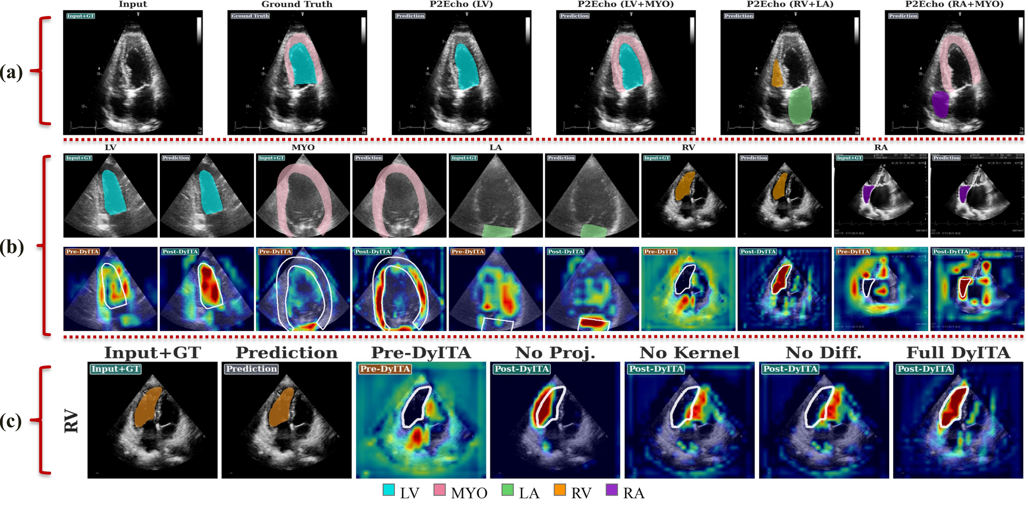
| Condition | Example prompt | Dice (%) ↑ | IoU (%) ↑ |
| Canonical | “Segment the LV: The LV is fully visualized from base to apex.” | 93.53 ± 6.83 | 88.39 ± 8.84 |
| Clinical paraphrase | “Delineate the left ventricular blood pool.” | 80.48 ± 23.88 | 72.62 ± 26.76 |
| Concise | “Segment the left ventricle cavity.” | 35.83 ± 36.23 | 28.58 ± 30.95 |
| Ambiguous | “Segment the cardiac chamber.” | 0.01 ± 0.23 | 0.01 ± 0.12 |
| Irrelevant | “Segment the liver.” | 6.35 ± 19.70 | 4.95 ± 16.54 |
| Null | “No anatomical structure is requested.” | 0.00 ± 0.00 | 0.00 ± 0.00 |
| Method | Time (s/image) ↓ |
| nnUNet | 0.04057 ± 0.00002 |
| SwinUNet | 0.00577 ± 0.00002 |
| MissFormer | 0.01034 ± 0.00001 |
| CENet | 0.02328 ± 0.00010 |
| SAMUS | 0.06620 ± 0.00024 |
| SAM3 (Zero-shot) | 0.19694 ± 0.00004 |
| SAM3 (Fine-tuned) | 0.08203 ± 0.00009 |
| MedCLIP-SAMv2 | 2.46376 ± 0.02072 |
| Grounding DINO-US-SAM | 2.59870 ± 0.01970 |
| EchoONE | 0.08649 ± 0.00004 |
| P2Echo (ours) | 0.01546 ± 0.00002 |
@article{heidari2026p2echo,
  title   = {Prompt2Echo (P2Echo): Text-Prompted Multi-View Echocardiography Segmentation with One Model},
  author  = {Heidari, Moein and Kimbowa, Alvin and Tabatabaei, Mahdi and Bozorgpour, Afshin and Ghorbani Kolahi, Sina and Jiang, River and Chen, Wenjin and Foran, David J. and Grewal, Jasmine and Merhof, Dorit and Hacihaliloglu, Ilker},
  journal = {arXiv preprint arXiv:ARXIV_ID_PLACEHOLDER},
  year    = {2026}
}