Moein Heidari

Moein Heidari

Pushing the boundaries of
AI in healthcare.

Ph.D. Candidate, Biomedical Engineering
University of British Columbia
UltrAi Lab

Explore my research

My research explores computer vision, medical imaging, and deep learning, with a focus on implicit neural representations, vision transformers, diffusion models, and AI agents and large language models for medical applications.

I am a Ph.D. candidate in Biomedical Engineering at the University of British Columbia, supervised by Dr. Ilker Hacihaliloglu at the UltrAi Lab. My thesis, Towards Complete AI-Enabled Echocardiography Interpretation with a Unified Multimodal Framework, combines multimodal AI agents with vision models to improve cardiac imaging analysis and diagnosis.

Previously, I was a remote research assistant at RWTH Aachen University with Prof. Dorit Merhof and Reza Azad, and at MBZUAI with Dr. Hisham Cholakkal. I received my M.Sc. in Communication Systems and B.Sc. in Electrical Engineering from the Iran University of Science and Technology.

I have collaborated with researchers at Stanford, RWTH Aachen, Mila, MBZUAI, and Rutgers, and I am always happy to discuss research ideas and collaborations.

View CV

Experience

Ph.D. in Biomedical Engineering

University of British Columbia, Vancouver, Canada

Jan. 2024 - Present

Research Assistant

Mohamed bin Zayed University of Artificial Intelligence (MBZUAI), Remote

Jan. 2023 - May 2023

AI Engineer

FaraAI, Tehran, Iran

May 2022 - Dec. 2022

Machine Learning and Computer Vision Researcher

DGSculptor, Montreal, Canada (Remote)

Dec. 2021 - Dec. 2023

Remote Research Assistant

RWTH Aachen University, Aachen, Germany

Oct. 2021 - Dec. 2023

M.Sc. in Communication Systems

Iran University of Science and Technology, Tehran, Iran

Oct. 2021 - Nov. 2023

Research Assistant

Iran University of Science and Technology, Dept. of Computer Science, Tehran, Iran

Apr. 2021 - Jul. 2021

B.Sc. in Electrical Engineering

Iran University of Science and Technology, Tehran, Iran

Sep. 2017 - Sep. 2021

Teaching

Teaching Assistant, DSCI 100: Introduction to Data Science

University of British Columbia

Fall 2025

Teaching Assistant, DSCI 430: Fairness, Accountability, Transparency and Ethics in Data Science

University of British Columbia

2025W2

Teaching Assistant, CPSC 330: Applied Machine Learning

University of British Columbia

2024W2

Teaching Assistant, CPSC 340: Machine Learning and Data Mining

University of British Columbia

2024W2

Teaching Assistant, Fundamentals of Deep Learning

Iran University of Science and Technology

Spring 2022

Teaching Assistant, Signals and Systems Analysis

Iran University of Science and Technology

Spring 2021, 2022, 2023

Talks

Research proposal: Towards Complete AI-Enabled Echocardiography Interpretation with a Unified Multimodal Framework

Ph.D. candidacy, University of British Columbia

Nov. 2025

Research presentation

Annual HLI Research Day

Aug. 2025

Service

Conference Reviewer

NeurIPS 2026 · ICML 2026 · ICLR 2024, 2025, 2026 · MICCAI 2024, 2025, 2026 · CVPR 2025 · ICCV 2025 · BMVC 2025, 2026

2024 - Present

Journal Reviewer

Transactions on Machine Learning Research (TMLR) · IEEE Transactions on Medical Imaging · IEEE Journal of Biomedical and Health Informatics · IEEE Access · IEEE Systems Journal · IEEE Signal Processing Letters

Honors & Awards

Impact Award, MIRASOL Workshop at MICCAI 2026, for WaRA

Oct. 2026

ICCV 2025 Broadening Participation Program award

Sep. 2025

Honorary master's admission, Iran University of Science and Technology

Sep. 2021

Ranked 3rd among 25 Communications students and 9th among 127 Electrical Engineering students, IUST

May 2021

1st place, national Rahneshan competition on detecting inappropriate content in images and videos (INEF)

Feb. 2021

Top 1% among ~148,000 participants in the Iranian National University Entrance Exam

2017

News

  • Nov. 2026

    🎉 Paper Which Pretext Task Transfers? Self-Supervised Pretraining Objectives for Lung Ultrasound accepted for an oral presentation at SPIE Medical Imaging 2027.

  • Oct. 2026

    🏆 Our paper WaRA won the Impact Award at the MIRASOL workshop at MICCAI 2026.

  • Oct. 2026

    🎉 Paper Incorporating Synthetic Data from Diffusion Models Supports Echocardiography Downstream Tasks published in the Journal of Medical Imaging.

  • Aug. 2026

    🎉 Four papers accepted at MICCAI 2026 workshops: Echo-E³Net (ASMUS), Echo-CoPilot (Agentic AI for Medicine), WaRA (MIRASOL, oral), and MaLViL (MLMI).

  • May 2026

    🎉 Paper XTinyU-Net: Training-Free U-Net Scaling via Initialization-Time Sensitivity received early acceptance at MICCAI 2026 (9% of more than 4,500 submissions).

  • Nov. 2025

    🎓 Successfully advanced to Ph.D. candidacy after presenting my research proposal, Towards Complete AI-Enabled Echocardiography Interpretation with a Unified Multimodal Framework.

  • Sep. 2025

    🏅 Selected for the ICCV 2025 Broadening Participation Program award to present my work at ICCV 2025 in Honolulu.

  • Aug. 2025

    🎤 Presented my research at the annual HLI Research Day.

  • Jun. 2025

    🎉 Paper SL²A-INR accepted at ICCV 2025.

  • Feb. 2025

    🎉 Paper Prompt2Perturb (P2P) accepted at CVPR 2025.

  • Dec. 2024

    🎉 Paper on Fourier Kolmogorov-Arnold Networks for implicit neural representations accepted at ICASSP 2025.

  • Jul. 2024

    🎉 Paper MSA²Net on multi-scale adaptive attention accepted at BMVC 2024.

  • Jan. 2024

    🎓 Started my Ph.D. in Biomedical Engineering at the University of British Columbia.

  • Aug. 2023

    🎉 Paper SA²-Net accepted for an oral presentation at BMVC 2023.

  • Aug. 2023

    🎉 Our survey on Vision Transformers in Medical Imaging accepted in Medical Image Analysis.

  • Aug. 2023

    🎉 Our survey on Diffusion Models in Medical Imaging accepted in Medical Image Analysis.

  • Sep. 2022

    🎉 Paper HiFormer on medical image segmentation accepted at WACV 2023.

  • Aug. 2022

    🎉 Three papers, Contextual Attention Network, IVD Labeling, and TransDeepLab, accepted at MICCAI 2022 workshops (MLMI and PRIME).

  • Aug. 2022

    🎉 Paper TransNorm accepted in IEEE Access.

  • Sep. 2021

    🎓 Defended my bachelor's thesis on deep learning based end-to-end wireless communication.

Publications

Browse the full list by year, then open individual abstracts, papers, code, and teaser visuals.

Showing all 27 publications.

Which Pretext Task Transfers? teaser

Which Pretext Task Transfers? Self-Supervised Pretraining Objectives for Lung Ultrasound

Moein Heidari, Junbo Rao, Jai Choraria, Wenjin Chen, David J. Foran, Ilker Hacihaliloglu

SPIE Medical Imaging 2027 | Oral presentation

paper code

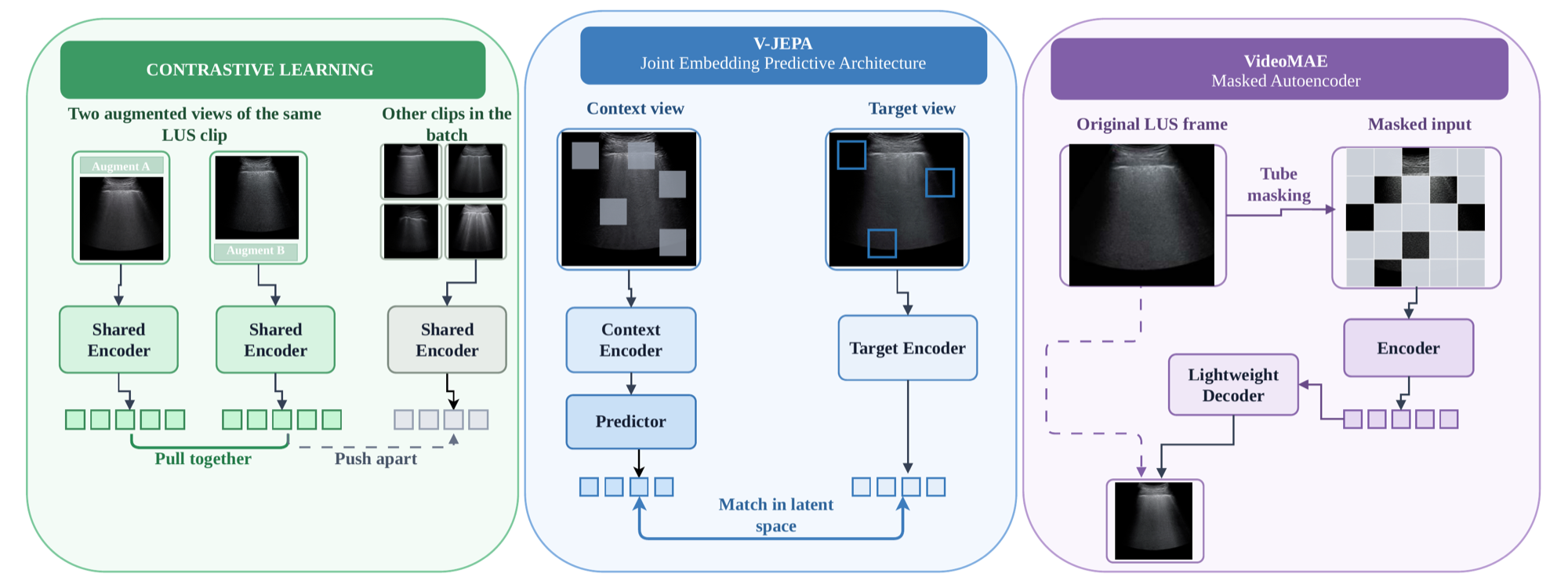
Self-supervised learning (SSL) can reduce the need for labelled medical images, but the choice of pretext objective remains unclear for lung ultrasound (LUS). Contrastive learning, masked reconstruction, and joint-embedding predictive architectures (JEPA) differ in the space in which their targets are defined, yet existing ultrasound studies compare them under different corpora, backbones, and evaluation protocols. We compare these three objective families using the same encoder backbone, pretraining corpus, optimisation schedule, and frozen-evaluation protocol. Encoders are pretrained on COVID-BLUeS LUS videos and evaluated with linear, kNN, and attentive probes at 5%, 10%, 50%, and 100% label budgets. Evaluation is performed on POCUS using patient-level five-fold cross-validation and on the independently acquired Mendeley-Uganda dataset, which is excluded from both pretraining and probe fitting. At the full label budget under linear probing, VideoMAE and V-JEPA achieve 66.5 ± 13.1 and 65.4 ± 11.7 balanced accuracy on POCUS, while MoCo achieves 42.1 ± 1.2. On Mendeley-Uganda, the ranking reverses: MoCo performs best at 62.7 ± 1.0, followed by VideoMAE at 53.8 ± 2.8, while V-JEPA falls near chance at 35.1 ± 4.9. These results show that POCUS probe accuracy alone does not identify the objective that transfers best across datasets. We also outline planned representation-level analyses to examine this reversal. Code is publicly available at https://github.com/moeinheidari7829/LUSVideoSSL.

Every Pixel Is an Option teaser

Every Pixel Is an Option: A System One Model for Medical Ultrasound Biometry

Moein Heidari, Junbo Rao, Yudan Chen, Yasamin Medghalchi, River Jiang, Wenjin Chen, David J. Foran, Jasmine Grewal, Ilker Hacihaliloglu

arXiv preprint, 2026

project paper

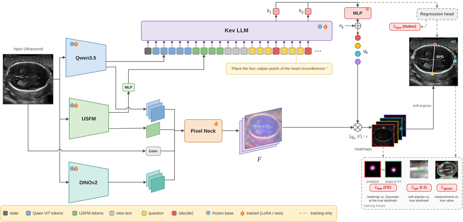
**Purpose:** Ultrasound biometry is widely used across various clinical applications, including prenatal and cardiac examinations, to obtain quantitative measurements by placing calipers at anatomical landmarks. However, these measurements remain subject to interoperator variability as they rely largely on manual caliper placement. Conventional methods often use specialized prediction heads for different ultrasound tasks, whereas vision-language models offer flexibility through text instructions. However, direct coordinate prediction does not explicitly model spatial likelihood distributions, limiting the representation of localization ambiguity. **Methods:** We propose Kev-Bio, a vision-language model that uses clinical measurement questions to guide ultrasound landmark detection. Inspired by System One decision models, Kev-Bio uses decision representations of the language model to generate queries for each caliper endpoint. These queries are matched against shared dense ultrasound features to produce spatial probability maps, from which landmark coordinates and clinical measurements are derived. The model is trained end-to-end using both landmark localization and clinical measurement losses. **Results:** On the nine-view FU/GU Biometry 2026 dataset, Kev-Bio achieves a mean radial error (MRE) of 22.50 pixels and a clinical measurement mean absolute error (MAE) of 15.38, outperforming the compared baselines in overall performance. **Conclusion:** These results demonstrate the effectiveness of Kev-Bio for ultrasound landmark detection and clinical measurement across different anatomical views. Our code and models are available at https://github.com/moeinheidari7829/Kev-Bio.
Synthetic echocardiography data (JMI) teaser

Incorporating Synthetic Data from Diffusion Models Supports Echocardiography Downstream Tasks

Milad Yazdani, Pooria Ashrafian, Moein Heidari, Alireza Rafiei Sardouei, Dena Shahriari, Ilker Hacihaliloglu

Journal of Medical Imaging (JMI), 2026

paper

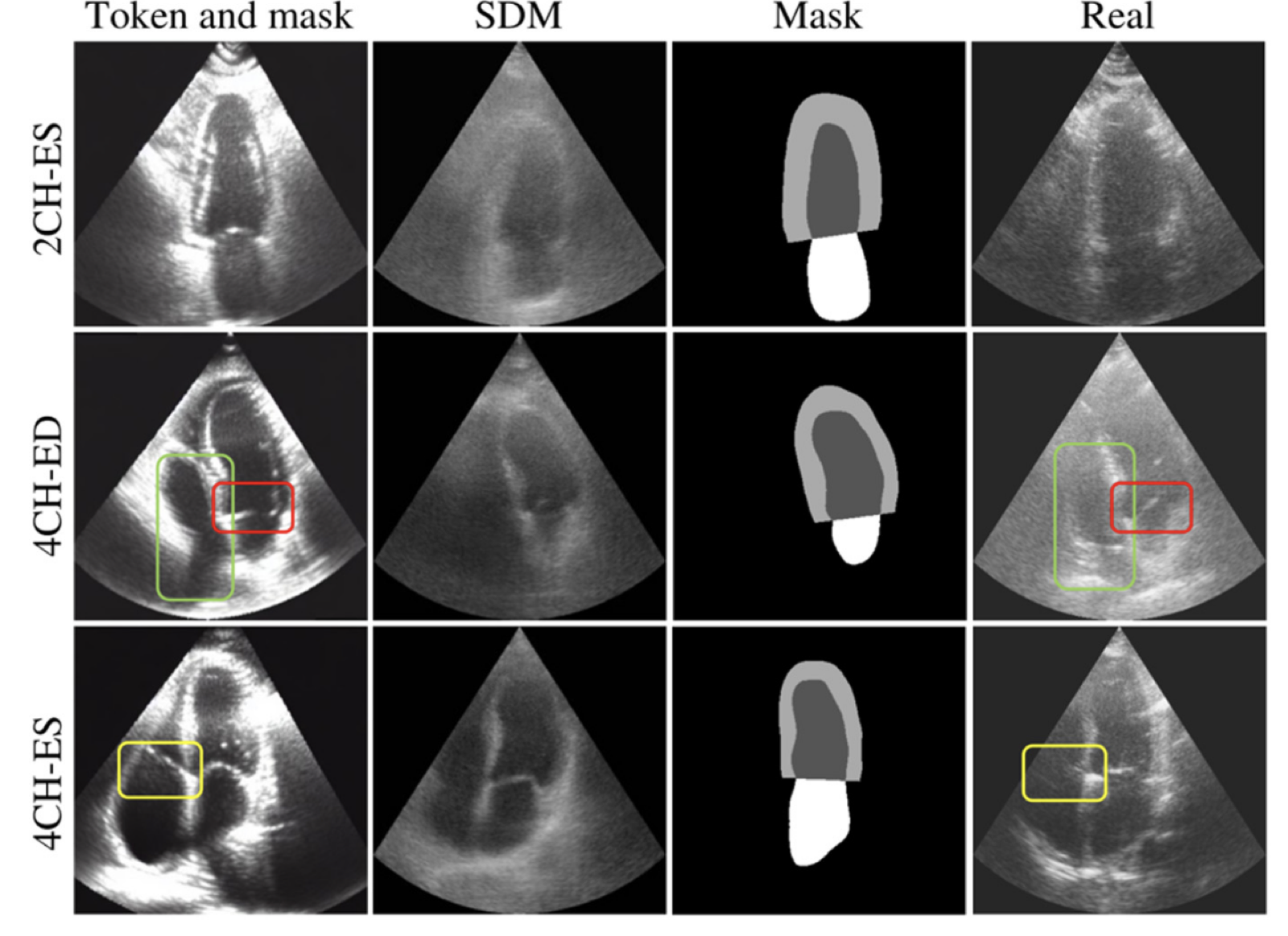
Purpose: Large labeled echocardiography datasets are costly to collect. We use diffusion models to generate synthetic training data for segmentation and cardiac phase classification. Approach: We study three generation approaches on CAMUS: an unconditional image space model, a latent model conditioned on plain English prompts or abstract tokens, and a ControlNet model conditioned on an abstract token and segmentation mask. We evaluate synthesis with Fréchet inception distance (FID) and augmentation using segmentation and classification of end diastole and end systole. We also evaluate segmentation within CardiacUDC and directly from CAMUS to CardiacUDC. Results: Conditioning on an abstract token and segmentation mask gives the lowest mean FID, 1.5, compared with 3.2 for the semantic diffusion model. Abstract token augmentation gives the highest classification accuracy, improving ResNet-18 from 83.8% to 87.2% and VGG-16 from 74.5% to 78.7%. Synthetic augmentation gives the highest mean Dice of 87.6% for the CAMUS 2D U-Net and 84.6% for the CardiacUDC nnU-Net. When nnU-Net models are trained on CAMUS and evaluated directly on CardiacUDC, augmentation raises mean Dice from 74.6% to 75.2%. Conclusions: Incorporating synthetic images into training gives modest gains in CAMUS phase classification, segmentation within CAMUS and CardiacUDC, and direct segmentation from CAMUS to CardiacUDC.

WaRA teaser

WaRA: Wavelet Low-Rank Adaptation for Medical Image Classification

Moein Heidari, Yijin Huang, Yasamin Medghalchi, Alireza Rafiei, Roger Tam, Ilker Hacihaliloglu

MICCAI 2026 MIRASOL Workshop | Oral presentation | Impact Award

project paper code

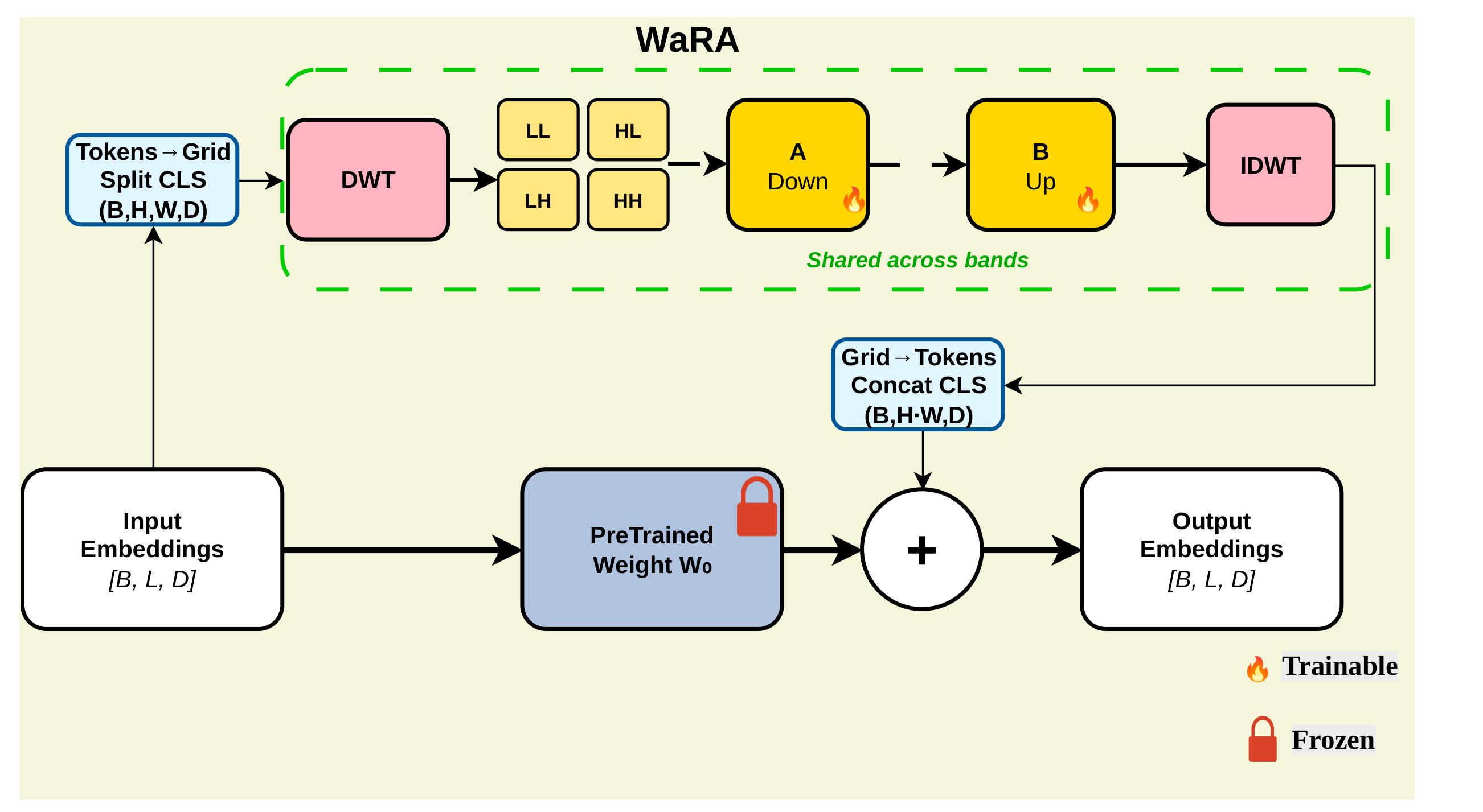
Adapting large pretrained vision models to medical image classification is often limited by memory, computation, and task-specific specializations. Parameter-efficient fine-tuning (PEFT) methods like LoRA reduce this cost by learning low-rank updates, but operating directly in feature space can struggle to capture the localized, multi-scale features common in medical imaging. We propose WaRA, a wavelet-structured adaptation module that performs low-rank adaptation in a wavelet domain. WaRA reshapes patch tokens into a spatial grid, applies a fixed discrete wavelet transform, updates subband coefficients using a shared low-rank adapter, and reconstructs the additive update through an inverse wavelet transform. This design provides a compact trainable interface while biasing the update toward both coarse structure and fine detail. For extremely low-resource settings, we introduce Tiny-WaRA, which further reduces trainable parameters by learning only a small set of coefficients in a fixed basis derived from the pretrained weights through a truncated SVD. Experiments on medical image classification across four modalities and datasets demonstrate that WaRA consistently improves performance over strong PEFT baselines, while retaining a favorable efficiency profile. Our code is publicly available at https://github.com/moeinheidari7829/WaRA.

Echo-CoPilot teaser

Echo-CoPilot: A Multiple-Perspective Agentic Framework for Reliable Echocardiography Interpretation

Moein Heidari, Ali Mehrabian, Mohammad Amin Roohi, River Jiang, Wenjin Chen, David J. Foran, Jasmine Grewal, Ilker Hacihaliloglu

MICCAI 2026 Agentic AI for Medicine Workshop

project paper code

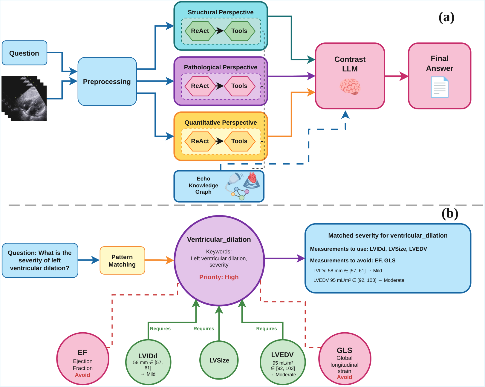
Echocardiography interpretation requires integrating multi-view temporal evidence with quantitative measurements and guideline-grounded reasoning, yet existing foundation-model pipelines largely solve isolated subtasks and fail when tool outputs are noisy or values fall near clinical cutoffs. We propose Echo-CoPilot, an end-to-end agentic framework that combines a multi-perspective workflow with knowledge-graph-guided measurement selection. Echo-CoPilot runs three independent ReAct-style agents, structural, pathological, and quantitative, that invoke specialized echocardiography tools to extract parameters while querying EchoKG to determine which measurements are required for the clinical question and which should be avoided. A self-contrast language model then compares the evidence-grounded perspectives, generates a discrepancy checklist, and re-queries EchoKG to apply the appropriate guideline thresholds and resolve conflicts, reducing hallucinated measurement selection and borderline flip-flops. On MIMICEchoQA, Echo-CoPilot provides higher accuracy compared to SOTA baselines and, under a stochasticity stress test, achieves higher reliability through more consistent conclusions and fewer answer changes across repeated runs. Our code is publicly available at https://github.com/moeinheidari7829/Echo-CoPilot.

XTinyU-Net teaser

XTinyU-Net: Training-Free U-Net Scaling via Initialization-Time Sensitivity

Alvin Kimbowa*, Moein Heidari*, David Liu, Ilker Hacihaliloglu
* Equal contribution

MICCAI 2026 (early accept)

paper code

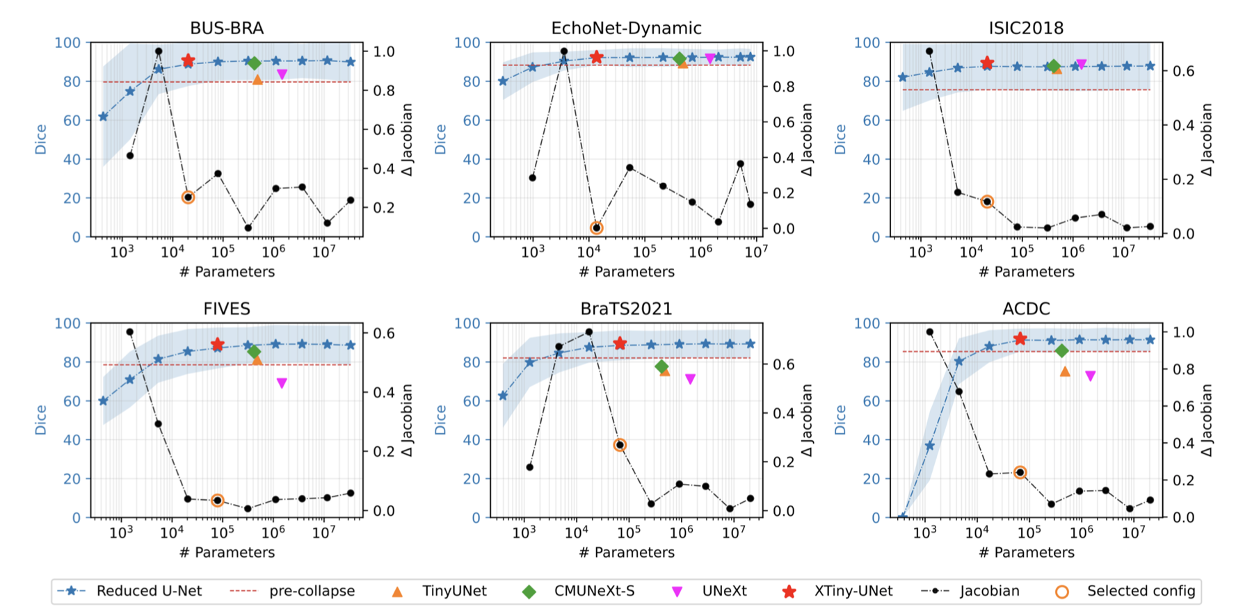
While U-Net architectures remain the gold standard for medical image segmentation, their deployment in resource-constrained environments demands aggressive model compression. However, finding an optimally efficient configuration is computationally prohibitive, typically requiring exhaustive train-and-evaluate cycles to find the smallest model that maintains peak performance. In this paper, we introduce a training-free selection framework to automatically identify ultralightweight, dataset-specific U-Net configurations directly at initialization. We observe that systematically scaling down U-Net channel width induces a sharp transition from a stable performance plateau to representational capacity collapse. To pinpoint this boundary without training, we propose a Jacobian-based sensitivity metric that scores discrete, width-capped U-Net variants using a small set of unlabeled images. By analyzing the total variation of this sensitivity curve, we isolate the smallest stable configuration, which we denote as XTinyU-Net. Evaluated across six diverse medical datasets within the nnU-Net framework, XTinyU-Net achieves segmentation accuracy comparable to the heavy nnU-Net baseline with 400x-1600x fewer parameters, and outperforms contemporary lightweight architectures while utilizing 5x-72x fewer parameters. Code is publicly accessible on https://github.com/alvinkimbowa/nntinyunet.git.

MaLViL teaser

MaLViL: Multi-axis Low-rank Vision-LSTM for Medical Image Segmentation

Afshin Bozorgpour, Sina Ghorbani Kolahi, Moein Heidari, Ilker Hacihaliloglu, Dorit Merhof

MICCAI 2026 MLMI Workshop

paper code

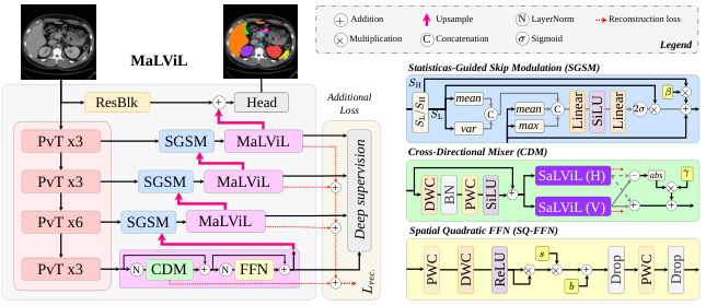
Vision-LSTM (ViL) enables efficient global modeling, but its cost still scales with the number of spatial tokens, so existing segmenters confine ViL to a coarse bottleneck and lose fine anatomical detail. Rasterizing 2D features into a 1D sequence further breaks adjacency across the orthogonal scan axis. We propose MaLViL, a Multi-axis Low-rank Vision-LSTM network that extends ViL across decoder resolutions. Bidirectional low-rank ViL (Bi-LRViL) reasons on a compact orthonormal subspace and preserves detail through an orthogonal residual; scale-aware SaLViL restores cross-axis neighbors before serialization; and a Cross-Directional Mixer (CDM) fuses orthogonal horizontal and vertical traversal paths. Statistics-Guided Skip Modulation (SGSM) further retains boundary cues in encoder skips. On skin-lesion, ultrasound, and multi-organ CT benchmarks, MaLViL achieves competitive or state-of-the-art segmentation accuracy, while reducing ViL operator memory by up to 83× at fine decoder resolutions. Code is available at: https://github.com/xmindflow/malvil.

Echo-E³Net teaser

Echo-E³Net: Efficient Endocardial Spatio-Temporal Network for Ejection Fraction Estimation

Moein Heidari*, Afshin Bozorgpour*, AmirHossein Zarif-Fakharnia, Wenjin Chen, Dorit Merhof, David J. Foran, Jasmine Grewal, Ilker Hacihaliloglu
* Equal contribution

MICCAI 2026 ASMUS Workshop

project paper code

Left ventricular ejection fraction (LVEF) is a primary marker of cardiac function. However, routine estimation from endocardial measurements requires manual delineation at end-diastole (ED) and end-systole (ES), a process that is time-consuming and subject to inter-observer variability. Reliable automation is especially valuable for point-of-care ultrasound (POCUS), where computational resources are limited and acquisition quality varies. We propose Echo-E³Net, an anatomy-guided spatio-temporal network that explicitly embeds cardiac anatomy into LVEF prediction. A dual-phase Endocardial Border Detector (E²CBD) uses phase-specific cross-attention to localize ED/ES endocardial landmarks and produce phase-aware landmark embeddings, while an Endocardial Feature Aggregator (E²FA) fuses these embeddings with global statistical descriptors of deep feature maps to refine EF regression. Training is guided by a lightweight geometric loss that uses ED and ES endocardial landmarks to regularize EF prediction. On EchoNet-Dynamic and a PSAX subset of EchoNet-Pediatric, Echo-E³Net attains competitive performance using only 1.55M parameters and 8.05 GFLOPs, an order-of-magnitude compute reduction versus recent baselines, supporting real-time deployment. Our code is publicly available at https://github.com/moeinheidari7829/Echo-E3Net.

Prompt2Perturb teaser

Prompt2Perturb (P2P): Text-Guided Diffusion-Based Adversarial Attacks on Breast Ultrasound Images

Yasamin Medghalchi, Moein Heidari, Clayton Allard, Leonid Sigal, Ilker Hacihaliloglu

CVPR 2025

paper code

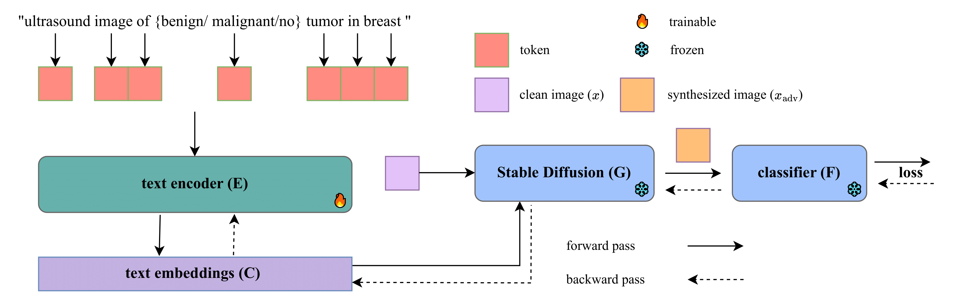
Deep neural networks (DNNs) offer significant promise for improving breast cancer diagnosis in medical imaging. However, these models are highly susceptible to adversarial attacks--small, imperceptible changes that can mislead classifiers--raising critical concerns about their reliability and security. Traditional attacks rely on fixed-norm perturbations, misaligning with human perception. In contrast, diffusion-based attacks require pre-trained models, demanding substantial data when these models are unavailable, limiting practical use in data-scarce scenarios. In medical imaging, however, this is often unfeasible due to the limited availability of datasets. Building on recent advancements in learnable prompts, we propose Prompt2Perturb (P2P), a novel language-guided attack method capable of generating meaningful attack examples driven by text instructions. During the prompt learning phase, our approach leverages learnable prompts within the text encoder to create subtle, yet impactful, perturbations that remain imperceptible while guiding the model towards targeted outcomes. In contrast to current prompt learning-based approaches, our P2P stands out by directly updating text embeddings, avoiding the need for retraining diffusion models. Further, we leverage the finding that optimizing only the early reverse diffusion steps boosts efficiency while ensuring that the generated adversarial examples incorporate subtle noise, thus preserving ultrasound image quality without introducing noticeable artifacts. We show that our method outperforms state-of-the-art attack techniques across three breast ultrasound datasets in FID and LPIPS. Moreover, the generated images are both more natural in appearance and more effective compared to existing adversarial attacks. Our code will be publicly available https://github.com/yasamin-med/P2P.

SL²A-INR teaser

SL²A-INR: Single-Layer Learnable Activation for Implicit Neural Representation

Moein Heidari*, Reza Rezaeian*, Reza Azad, Dorit Merhof, Hamid Soltanian-Zadeh, Ilker Hacihaliloglu
* Equal contribution

ICCV 2025

project paper code video

Implicit Neural Representation (INR), leveraging a neural network to transform coordinate input into corresponding attributes, has recently driven significant advances in several vision-related domains. However, the performance of INR is heavily influenced by the choice of the nonlinear activation function used in its multilayer perceptron (MLP) architecture. To date, multiple nonlinearities have been investigated, but current INRs still face limitations in capturing high-frequency components and diverse signal types. We show that these challenges can be alleviated by introducing a novel approach in INR architecture. Specifically, we propose SL²A-INR, a hybrid network that combines a single-layer learnable activation function with an MLP that uses traditional ReLU activations. Our method performs superior across diverse tasks, including image representation, 3D shape reconstruction, and novel view synthesis. Through comprehensive experiments, SL²A-INR sets new benchmarks in accuracy, quality, and robustness for INR. Our Code is publicly available on GitHub.

Fourier KAN teaser

Implicit Neural Representations with Fourier Kolmogorov-Arnold Networks

Ali Mehrabian, Parsa Mojarad Adi, Moein Heidari, Ilker Hacihaliloglu

ICASSP 2025

paper code

Implicit neural representations (INRs) use neural networks to provide continuous and resolution-independent representations of complex signals with a small number of parameters. However, existing INR models often fail to capture important frequency components specific to each task. To address this issue, in this paper, we propose a Fourier Kolmogorov Arnold network (FKAN) for INRs. The proposed FKAN utilizes learnable activation functions modeled as Fourier series in the first layer to effectively control and learn the task-specific frequency components. In addition, the activation functions with learnable Fourier coefficients improve the ability of the network to capture complex patterns and details, which is beneficial for high-resolution and high-dimensional data. Experimental results show that our proposed FKAN model outperforms three state-of-the-art baseline schemes, and improves the peak signal-to-noise ratio (PSNR) and structural similarity index measure (SSIM) for the image representation task and intersection over union (IoU) for the 3D occupancy volume representation task, respectively. The code is available at github.com/Ali-Meh619/FKAN.

U-Net modifications for retroperitoneal tumor segmentation teaser

A Study on the Performance of U-Net Modifications in Retroperitoneal Tumor Segmentation

Moein Heidari, Ehsan Khodapanah Aghdam, Alexander Manzella, Daniel Hsu, Rebecca Scalabrino, Wenjin Chen, David J. Foran, Ilker Hacihaliloglu

SPIE Medical Imaging 2025

project paper code

The retroperitoneum hosts a variety of tumors, including rare benign and malignant types, which pose diagnostic and treatment challenges due to their infrequency and proximity to vital structures. Estimating tumor volume is difficult due to their irregular shapes, and manual segmentation is time-consuming. Automatic segmentation using U-Net and its variants, incorporating Vision Transformer (ViT) elements, has shown promising results but struggles with high computational demands. To address this, architectures like the Mamba State Space Model (SSM) and Extended Long-Short Term Memory (xLSTM) offer efficient solutions by handling long-range dependencies with lower resource consumption. This study evaluates U-Net enhancements, including CNN, ViT, Mamba, and xLSTM, on a new in-house CT dataset and a public organ segmentation dataset. The proposed ViLU-Net model integrates Vi-blocks for improved segmentation. Results highlight xLSTM's efficiency in the U-Net framework. The code is publicly accessible on GitHub.

MSA²Net teaser

MSA²Net: Multi-scale Adaptive Attention-guided Network for Medical Image Segmentation

Sina Ghorbani Kolahi, Seyed Kamal Chaharsooghi, Toktam Khatibi, Afshin Bozorgpour, Reza Azad, Moein Heidari, Ilker Hacihaliloglu, Dorit Merhof

BMVC 2024

paper code

Medical image segmentation involves identifying and separating object instances in a medical image to delineate various tissues and structures, a task complicated by the significant variations in size, shape, and density of these features. Convolutional neural networks (CNNs) have traditionally been used for this task but have limitations in capturing long-range dependencies. Transformers, equipped with self-attention mechanisms, aim to address this problem. However, in medical image segmentation it is beneficial to merge both local and global features to effectively integrate feature maps across various scales, capturing both detailed features and broader semantic elements for dealing with variations in structures. In this paper, we introduce MSA²Net, a new deep segmentation framework featuring an expedient design of skip-connections. These connections facilitate feature fusion by dynamically weighting and combining coarse-grained encoder features with fine-grained decoder feature maps. Specifically, we propose a Multi-Scale Adaptive Spatial Attention Gate (MASAG), which dynamically adjusts the receptive field (Local and Global contextual information) to ensure that spatially relevant features are selectively highlighted while minimizing background distractions. Extensive evaluations involving dermatology, and radiological datasets demonstrate that our MSA²Net outperforms state-of-the-art (SOTA) works or matches their performance. The source code is publicly available at https://github.com/xmindflow/MSA-2Net.

State space models survey teaser

Computation-Efficient Era: A Comprehensive Survey of State Space Models in Medical Image Analysis

Moein Heidari, Sina Ghorbani Kolahi, Sanaz Karimijafarbigloo, Bobby Azad, Afshin Bozorgpour, Soheila Hatami, Reza Azad, Ali Diba, Ulas Bagci, Dorit Merhof

arXiv preprint, 2024

project paper code

Sequence modeling plays a vital role across various domains, with recurrent neural networks being historically the predominant method of performing these tasks. However, the emergence of transformers has altered this paradigm due to their superior performance. Built upon these advances, transformers have conjoined CNNs as two leading foundational models for learning visual representations. However, transformers are hindered by the O(N²) complexity of their attention mechanisms, while CNNs lack global receptive fields and dynamic weight allocation. State Space Models (SSMs), specifically the Mamba model with selection mechanisms and hardware-aware architecture, have garnered immense interest lately in sequential modeling and visual representation learning, challenging the dominance of transformers by providing infinite context lengths and offering substantial efficiency maintaining linear complexity in the input sequence. Capitalizing on the advances in computer vision, medical imaging has heralded a new epoch with Mamba models. Intending to help researchers navigate the surge, this survey seeks to offer an encyclopedic review of Mamba models in medical imaging. Specifically, we start with a comprehensive theoretical review forming the basis of SSMs, including Mamba architecture and its alternatives for sequence modeling paradigms in this context. Next, we offer a structured classification of Mamba models in the medical field and introduce a diverse categorization scheme based on their application, imaging modalities, and targeted organs. Finally, we summarize key challenges, discuss different future research directions of the SSMs in the medical domain, and propose several directions to fulfill the demands of this field. In addition, we have compiled the studies discussed in this paper along with their open-source implementations on our GitHub repository.

Efficient vision transformers teaser

Enhancing Efficiency in Vision Transformer Networks: Design Techniques and Insights

Moein Heidari, Reza Azad, Sina Ghorbani Kolahi, René Arimond, Leon Niggemeier, Alaa Sulaiman, Afshin Bozorgpour, Ehsan Khodapanah Aghdam, Amirhossein Kazerouni, Ilker Hacihaliloglu, Dorit Merhof

arXiv preprint, 2024

project paper

Intrigued by the inherent ability of the human visual system to identify salient regions in complex scenes, attention mechanisms have been seamlessly integrated into various Computer Vision (CV) tasks. Building upon this paradigm, Vision Transformer (ViT) networks exploit attention mechanisms for improved efficiency. This review navigates the landscape of redesigned attention mechanisms within ViTs, aiming to enhance their performance. This paper provides a comprehensive exploration of techniques and insights for designing attention mechanisms, systematically reviewing recent literature in the field of CV. This survey begins with an introduction to the theoretical foundations and fundamental concepts underlying attention mechanisms. We then present a systematic taxonomy of various attention mechanisms within ViTs, employing redesigned approaches. A multi-perspective categorization is proposed based on their application, objectives, and the type of attention applied. The analysis includes an exploration of the novelty, strengths, weaknesses, and an in-depth evaluation of the different proposed strategies. This culminates in the development of taxonomies that highlight key properties and contributions. Finally, we gather the reviewed studies along with their available open-source implementations at our GitHub. We aim to regularly update it with the most recent relevant papers.

Vision-language synthetic echocardiography teaser

Vision-Language Synthetic Data Enhances Echocardiography Downstream Tasks

Pooria Ashrafian, Milad Yazdani, Moein Heidari, Dena Shahriari, Ilker Hacihaliloglu

arXiv preprint, 2024

paper code

High-quality, large-scale data is essential for robust deep learning models in medical applications, particularly ultrasound image analysis. Diffusion models facilitate high-fidelity medical image generation, reducing the costs associated with acquiring and annotating new images. This paper utilizes recent vision-language models to produce diverse and realistic synthetic echocardiography image data, preserving key features of the original images guided by textual and semantic label maps. Specifically, we investigate three potential avenues: unconditional generation, generation guided by text, and a hybrid approach incorporating both textual and semantic supervision. We show that the rich contextual information present in the synthesized data potentially enhances the accuracy and interpretability of downstream tasks, such as echocardiography segmentation and classification with improved metrics and faster convergence. Our implementation with checkpoints, prompts, and the created synthetic dataset will be publicly available at GitHub.

Vision transformers review teaser

Advances in Medical Image Analysis with Vision Transformers: A Comprehensive Review

Reza Azad, Amirhossein Kazerouni, Moein Heidari, Ehsan Khodapanah Aghdam, Amirali Molaei, Yiwei Jia, Abin Jose, Rijo Roy, Dorit Merhof

Medical Image Analysis (MedIA), 2024

paper code

The remarkable performance of the Transformer architecture in natural language processing has recently also triggered broad interest in Computer Vision. Among other merits, Transformers are witnessed as capable of learning long-range dependencies and spatial correlations, which is a clear advantage over convolutional neural networks (CNNs), which have been the de facto standard in Computer Vision problems so far. Thus, Transformers have become an integral part of modern medical image analysis. In this review, we provide an encyclopedic review of the applications of Transformers in medical imaging. Specifically, we present a systematic and thorough review of relevant recent Transformer literature for different medical image analysis tasks, including classification, segmentation, detection, registration, synthesis, and clinical report generation. For each of these applications, we investigate the novelty, strengths and weaknesses of the different proposed strategies and develop taxonomies highlighting key properties and contributions. Further, if applicable, we outline current benchmarks on different datasets. Finally, we summarize key challenges and discuss different future research directions. In addition, we have provided cited papers with their corresponding implementations in https://github.com/mindflow-institue/Awesome-Transformer.

SA²-Net teaser

SA²-Net: Scale-aware Attention Network for Microscopic Image Segmentation

Mustansar Fiaz, Moein Heidari, Rao Muhammad Anwer, Hisham Cholakkal

BMVC 2023 | Oral presentation

paper code

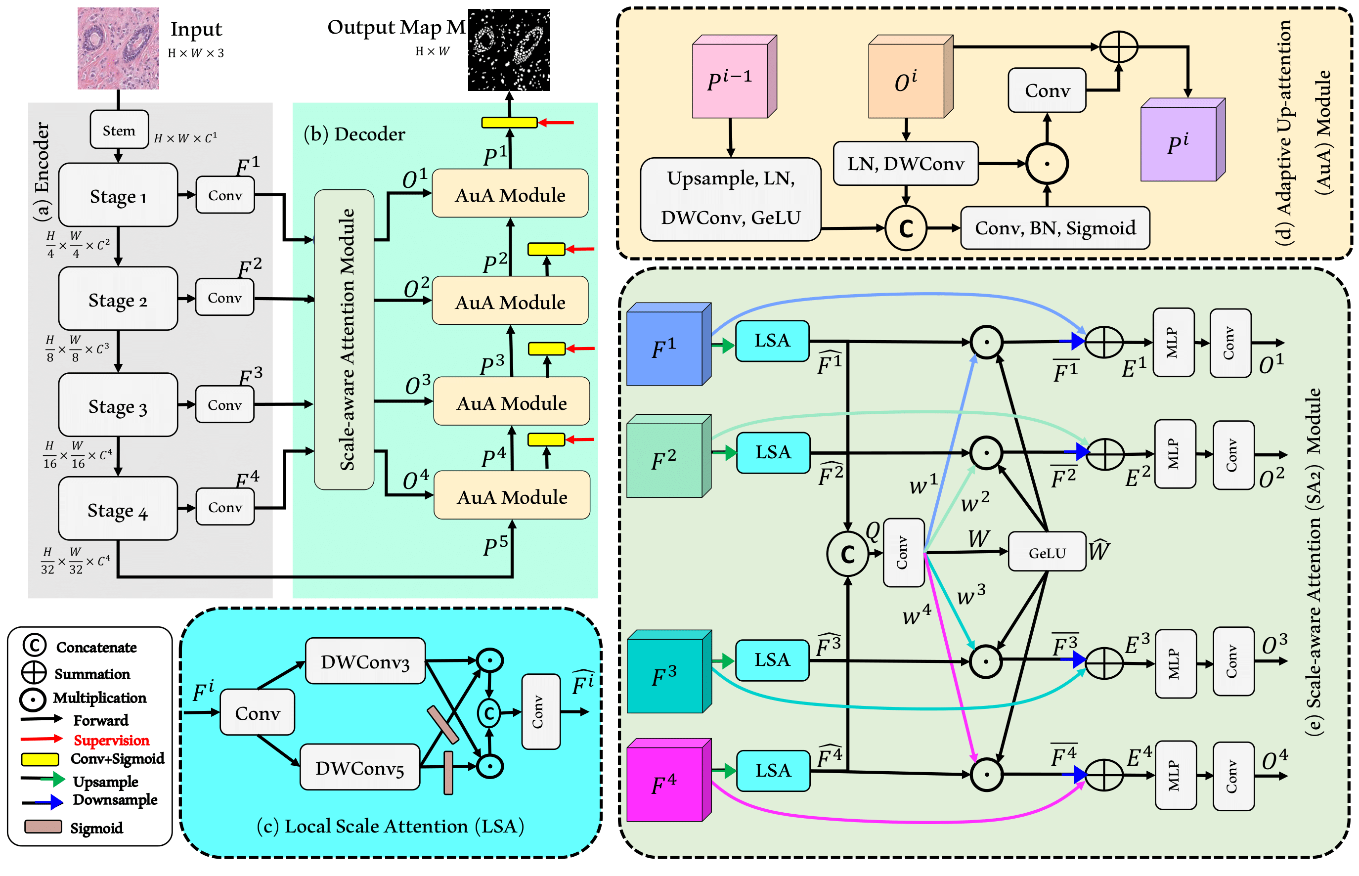
Microscopic image segmentation is a challenging task, wherein the objective is to assign semantic labels to each pixel in a given microscopic image. While convolutional neural networks (CNNs) form the foundation of many existing frameworks, they often struggle to explicitly capture long-range dependencies. Although transformers were initially devised to address this issue using self-attention, it has been proven that both local and global features are crucial for addressing diverse challenges in microscopic images, including variations in shape, size, appearance, and target region density. In this paper, we introduce SA2-Net, an attention-guided method that leverages multi-scale feature learning to effectively handle diverse structures within microscopic images. Specifically, we propose scale-aware attention (SA2) module designed to capture inherent variations in scales and shapes of microscopic regions, such as cells, for accurate segmentation. This module incorporates local attention at each level of multi-stage features, as well as global attention across multiple resolutions. Furthermore, we address the issue of blurred region boundaries (e.g., cell boundaries) by introducing a novel upsampling strategy called the Adaptive Up-Attention (AuA) module. This module enhances the discriminative ability for improved localization of microscopic regions using an explicit attention mechanism. Extensive experiments on five challenging datasets demonstrate the benefits of our SA2-Net model. Our source code is publicly available at https://github.com/mustansarfiaz/SA2-Net.

HiFormer teaser

HiFormer: Hierarchical Multi-scale Representations Using Transformers for Medical Image Segmentation

Moein Heidari*, Amirhossein Kazerouni*, Milad Soltany*, Reza Azad, Ehsan Khodapanah Aghdam, Julien Cohen-Adad, Dorit Merhof
* Equal contribution

WACV 2023

project paper code

Convolutional neural networks (CNNs) have been the consensus for medical image segmentation tasks. However, they suffer from the limitation in modeling long-range dependencies and spatial correlations due to the nature of convolution operation. Although transformers were first developed to address this issue, they fail to capture low-level features. In contrast, it is demonstrated that both local and global features are crucial for dense prediction, such as segmenting in challenging contexts. In this paper, we propose HiFormer, a novel method that efficiently bridges a CNN and a transformer for medical image segmentation. Specifically, we design two multi-scale feature representations using the seminal Swin Transformer module and a CNN-based encoder. To secure a fine fusion of global and local features obtained from the two aforementioned representations, we propose a Double-Level Fusion (DLF) module in the skip connection of the encoder-decoder structure. Extensive experiments on various medical image segmentation datasets demonstrate the effectiveness of HiFormer over other CNN-based, transformer-based, and hybrid methods in terms of computational complexity, and quantitative and qualitative results. Our code is publicly available at: https://github.com/amirhossein-kz/HiFormer

Diffusion models survey teaser

Diffusion Models in Medical Imaging: A Comprehensive Survey

Amirhossein Kazerouni, Ehsan Khodapanah Aghdam, Moein Heidari, Reza Azad, Mohsen Fayyaz, Ilker Hacihaliloglu, Dorit Merhof

Medical Image Analysis (MedIA), 2023

paper code

Denoising diffusion models, a class of generative models, have garnered immense interest lately in various deep-learning problems. A diffusion probabilistic model defines a forward diffusion stage where the input data is gradually perturbed over several steps by adding Gaussian noise and then learns to reverse the diffusion process to retrieve the desired noise-free data from noisy data samples. Diffusion models are widely appreciated for their strong mode coverage and quality of the generated samples despite their known computational burdens. Capitalizing on the advances in computer vision, the field of medical imaging has also observed a growing interest in diffusion models. To help the researcher navigate this profusion, this survey intends to provide a comprehensive overview of diffusion models in the discipline of medical image analysis. Specifically, we introduce the solid theoretical foundation and fundamental concepts behind diffusion models and the three generic diffusion modelling frameworks: diffusion probabilistic models, noise-conditioned score networks, and stochastic differential equations. Then, we provide a systematic taxonomy of diffusion models in the medical domain and propose a multi-perspective categorization based on their application, imaging modality, organ of interest, and algorithms. To this end, we cover extensive applications of diffusion models in the medical domain. Furthermore, we emphasize the practical use case of some selected approaches, and then we discuss the limitations of the diffusion models in the medical domain and propose several directions to fulfill the demands of this field. Finally, we gather the overviewed studies with their available open-source implementations at https://github.com/amirhossein-kz/Awesome-Diffusion-Models-in-Medical-Imaging.

DiffGANPaint teaser

DiffGANPaint: Fast Inpainting Using Denoising Diffusion GANs

Moein Heidari, Alireza Morsali, Tohid Abedini, Samin Heydarian

ICLR 2023 Tiny Papers

project paper

Free-form image inpainting is the task of reconstructing parts of an image specified by an arbitrary binary mask. In this task, it is typically desired to generalize model capabilities to unseen mask types, rather than learning certain mask distributions. Capitalizing on the advances in diffusion models, in this paper, we propose a Denoising Diffusion Probabilistic Model (DDPM) based model capable of filling missing pixels fast as it models the backward diffusion process using the generator of a generative adversarial network (GAN) network to reduce sampling cost in diffusion models. Experiments on general-purpose image inpainting datasets verify that our approach performs superior or on par with most contemporary works.

MLP-Attention teaser

MLP-Attention: Improving Transformer Architecture with MLP Attention Weights

Alireza Morsali, Moein Heidari, Samin Heydarian, Tohid Abedini

ICLR 2023 Tiny Papers

paper code

The Transformer architecture has revolutionized natural language processing (NLP) and has achieved state-of-the-art results in various tasks. The attention mechanism is one of the key components of the Transformer architecture, which allows the model to focus on relevant parts of the input. In the standard Transformer, the attention weights are computed by the dot product of query and key vectors followed by a softmax function. However, in this paper, we propose to replace the dot product of query and key vectors with a multi-layer perceptron (MLP) to compute attention weights directly from the embeddings. The proposed modification is simple and can be easily implemented in existing Transformer-based models to improve their performance as shown in this paper for an NLP task. We provide the implementation code at https://github.com/AlirezaMorsali/MLP-Attention for reproducibility and ease of adoption.

Segmentation loss functions survey teaser

Loss Functions in the Era of Semantic Segmentation: A Survey and Outlook

Reza Azad, Moein Heidari, Kadir Yilmaz, Michael Hüttemann, Sanaz Karimijafarbigloo, Yuli Wu, Anke Schmeink, Dorit Merhof

arXiv preprint, 2023

paper code

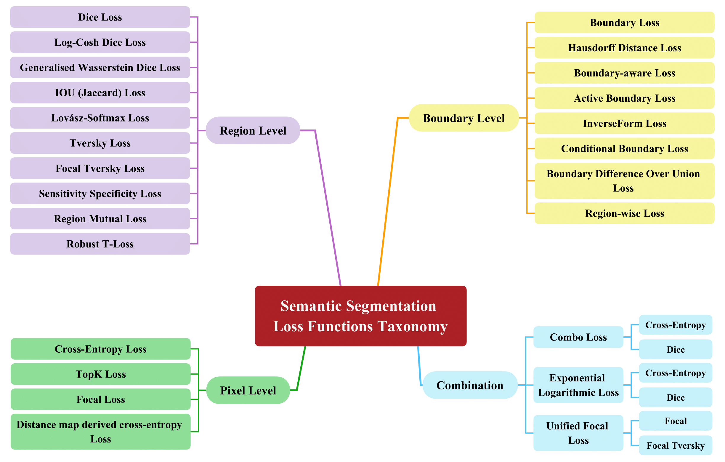
Semantic image segmentation, the process of classifying each pixel in an image into a particular class, plays an important role in many visual understanding systems. As the predominant criterion for evaluating the performance of statistical models, loss functions are crucial for shaping the development of deep learning-based segmentation algorithms and improving their overall performance. To aid researchers in identifying the optimal loss function for their particular application, this survey provides a comprehensive and unified review of 25 loss functions utilized in image segmentation. We provide a novel taxonomy and thorough review of how these loss functions are customized and leveraged in image segmentation, with a systematic categorization emphasizing their significant features and applications. Furthermore, to evaluate the efficacy of these methods in real-world scenarios, we propose unbiased evaluations of some distinct and renowned loss functions on established medical and natural image datasets. We conclude this review by identifying current challenges and unveiling future research opportunities. Finally, we have compiled the reviewed studies that have open-source implementations on our GitHub page.

Contextual Attention Network teaser

Contextual Attention Network: Transformer Meets U-Net

Reza Azad, Moein Heidari, Yuli Wu, Dorit Merhof

MICCAI 2022 MLMI Workshop

paper

Currently, convolutional neural networks (CNN) (e.g., U-Net) have become the de facto standard and attained immense success in medical image segmentation. However, as a downside, CNN based methods are a double-edged sword as they fail to build long-range dependencies and global context connections due to the limited receptive field that stems from the intrinsic characteristics of the convolution operation. Hence, recent articles have exploited Transformer variants for medical image segmentation tasks which open up great opportunities due to their innate capability of capturing long-range correlations through the attention mechanism. Although being feasibly designed, most of the cohort studies incur prohibitive performance in capturing local information, thereby resulting in less lucidness of boundary areas. In this paper, we propose a contextual attention network to tackle the aforementioned limitations. The proposed method uses the strength of the Transformer module to model the long-range contextual dependency. Simultaneously, it utilizes the CNN encoder to capture local semantic information. In addition, an object-level representation is included to model the regional interaction map. The extracted hierarchical features are then fed to the contextual attention module to adaptively recalibrate the representation space using the local information. Then, they emphasize the informative regions while taking into account the long-range contextual dependency derived by the Transformer module. We validate our method on several large-scale public medical image segmentation datasets and achieve state-of-the-art performance. We have provided the implementation code in https://github.com/rezazad68/TMUnet.

Intervertebral disc labeling teaser

Intervertebral Disc Labeling With Learning Shape Information, A Look Once Approach

Reza Azad, Moein Heidari, Julien Cohen-Adad, Ehsan Adeli, Dorit Merhof

MICCAI 2022 PRIME Workshop

paper

Accurate and automatic segmentation of intervertebral discs from medical images is a critical task for the assessment of spine-related diseases such as osteoporosis, vertebral fractures, and intervertebral disc herniation. To date, various approaches have been developed in the literature which routinely relies on detecting the discs as the primary step. A disadvantage of many cohort studies is that the localization algorithm also yields false-positive detections. In this study, we aim to alleviate this problem by proposing a novel U-Net-based structure to predict a set of candidates for intervertebral disc locations. In our design, we integrate the image shape information (image gradients) to encourage the model to learn rich and generic geometrical information. This additional signal guides the model to selectively emphasize the contextual representation and suppress the less discriminative features. On the post-processing side, to further decrease the false positive rate, we propose a permutation invariant 'look once' model, which accelerates the candidate recovery procedure. In comparison with previous studies, our proposed approach does not need to perform the selection in an iterative fashion. The proposed method was evaluated on the spine generic public multi-center dataset and demonstrated superior performance compared to previous work. We have provided the implementation code in https://github.com/rezazad68/intervertebral-lookonce

TransDeepLab teaser

TransDeepLab: Convolution-Free Transformer-based DeepLab v3+ for Medical Image Segmentation

Reza Azad, Moein Heidari, Moein Shariatnia, Ehsan Khodapanah Aghdam, Sanaz Karimijafarbigloo, Ehsan Adeli, Dorit Merhof

MICCAI 2022 PRIME Workshop

paper

Convolutional neural networks (CNNs) have been the de facto standard in a diverse set of computer vision tasks for many years. Especially, deep neural networks based on seminal architectures such as U-shaped models with skip-connections or atrous convolution with pyramid pooling have been tailored to a wide range of medical image analysis tasks. The main advantage of such architectures is that they are prone to detaining versatile local features. However, as a general consensus, CNNs fail to capture long-range dependencies and spatial correlations due to the intrinsic property of confined receptive field size of convolution operations. Alternatively, Transformer, profiting from global information modelling that stems from the self-attention mechanism, has recently attained remarkable performance in natural language processing and computer vision. Nevertheless, previous studies prove that both local and global features are critical for a deep model in dense prediction, such as segmenting complicated structures with disparate shapes and configurations. To this end, this paper proposes TransDeepLab, a novel DeepLab-like pure Transformer for medical image segmentation. Specifically, we exploit hierarchical Swin-Transformer with shifted windows to extend the DeepLabv3 and model the Atrous Spatial Pyramid Pooling (ASPP) module. A thorough search of the relevant literature yielded that we are the first to model the seminal DeepLab model with a pure Transformer-based model. Extensive experiments on various medical image segmentation tasks verify that our approach performs superior or on par with most contemporary works on an amalgamation of Vision Transformer and CNN-based methods, along with a significant reduction of model complexity. The codes and trained models are publicly available at https://github.com/rezazad68/transdeeplab

TransNorm teaser

TransNorm: Transformer Provides a Strong Spatial Normalization Mechanism for a Deep Segmentation Model

Reza Azad, Mohammad T. AL-Antary, Moein Heidari, Dorit Merhof

IEEE Access, 2022

paper

In the past few years, convolutional neural networks (CNNs), particularly U-Net, have been the prevailing technique in the medical image processing era. Specifically, the seminal U-Net, as well as its alternatives, have successfully managed to address a wide variety of medical image segmentation tasks. However, these architectures are intrinsically imperfect as they fail to exhibit long-range interactions and spatial dependencies leading to a severe performance drop in the segmentation of medical images with variable shapes and structures. Transformers, preliminary proposed for sequence-to-sequence prediction, have arisen as surrogate architectures to precisely model global information assisted by the self-attention mechanism. Despite being feasibly designed, utilizing a pure Transformer for image segmentation purposes can result in limited localization capacity stemming from inadequate low-level features. Thus, a line of research strives to design robust variants of Transformer-based U-Net. In this paper, we propose Trans-Norm, a novel deep segmentation framework which concomitantly consolidates a Transformer module into both encoder and skip-connections of the standard U-Net. We argue that the expedient design of skip-connections can be crucial for accurate segmentation as it can assist in feature fusion between the expanding and contracting paths. In this respect, we derive a Spatial Normalization mechanism from the Transformer module to adaptively recalibrate the skip connection path. Extensive experiments across three typical tasks for medical image segmentation demonstrate the effectiveness of TransNorm. The codes and trained models are publicly available at https://github.com/rezazad68/transnorm.

Contact

I'm always interested in discussing research ideas, collaborations, and opportunities. Feel free to reach out.